Posts

Complement Pathways: Types, Functions and Regulation

Image
Complement Pathways: Types, Functions and Regulation Complement was discovered by Jules Bordet as a heat-labile component of normal plasma that causes the opsonisation and killing of bacteria. It helps antibodies and phagocytic cells to clear pathogens and damaged cells; promote inflammation and attack pathogen’s plasma membrane. Proteins that take part in the complement system are called complements that collectively work as a biological cascade The complement system refers to a series of >20 proteins, circulating in the blood and tissue fluids. Most of the proteins are normally inactive, but in response to the recognition of molecular components of microorganisms they become sequentially activated in an enzyme cascade – the activation of one protein enzymatically cleaves and activates the next protein in the cascade. Complements are mainly denoted by the capital letter C with numbers; like, C1, C2, C3, and so on. Some have only alphabet, like, B , D . Some are simpl

Bacteriology - Flagellar Movement

Image
Flagellar Movement                   F ive major methods of movement have been observed: swimming movement conferred by flagella, flagella-mediated swarming, corkscrew movement of spirochetes, twitching motility associated with type IV pili, and gliding motility.                                   M otile bacteria do not move aimlessly. Rather, motility is used to move toward nutrients such as sugars and amino acids and away from many harmful substances and bacterial waste products.                                    M ovement toward chemical attractants and away from repellents is known as chemotaxis . Motile bacteria also can move in response to environmental cues such as temperature (thermotaxis), light (phototaxis), oxygen (aerotaxis), osmotic pressure (osmotaxis), and gravity.flagellar rotation results in two types of movement: a smooth swimming movement often called a run, which actually moves the cell from one spot to another, and a tumble, which serves to reorien

Bacteriology - Bacterial Flagella: structure, types and function

Image
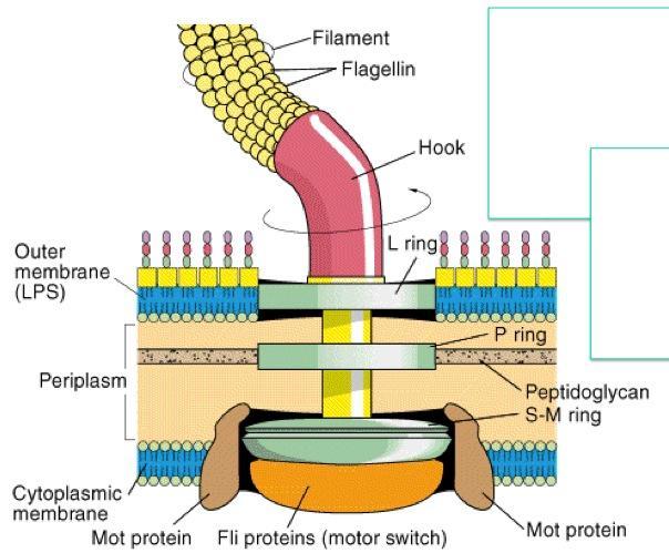
Bacterial Flagella: structure, types and function Many motile bacteria move by use of flagella (s., flagellum), threadlike locomotor appendages extending outward from the plasma membrane and cell wall. OR A flagellum (plural: flagella ) is a long, whip-like structure that helps some single celled organisms move. The synthesis of bacterial flagella is complex and involves at least 20 to 30 genes. Besides the gene for flagellin, 10 or more genes code for hook and basal body proteins; other genes are concerned with control of flagellar construction or function. Both prokaryotic and eukaryotic cells contain cilia and flagella Cilia and flagella are formed from specialized groupings of microtubules called basal bodies. If the protrusions are short and numerous they are termed cilia . If they are longer and less numerous (usually only one or two) they are termed flagella .  Although the main function of flagella is motility, they can have other roles. They can be involved in

Glycocalyx - Definition,Function

Image
Glycocalyx A layer of carbohydrate on the surface of the plasma membrane of most eukaryotic cells. It is made up of the oligosaccharide side-chains of the glycolipid and glycoprotein components of the membrane and may include oligosaccharides secreted by the cell. It plays a role in cell–cell adhesion and in regulating the exchange of materials between a cell and its environment. The glycocalyx is a carbohydrate-enriched coating that covers the outside of many eukaryotic cells and prokaryotic cells, particularly bacteria . On bacterial cells, the glycocalyx provides a protective coat from host factors. The outermost layer of a bacterium, typically consisting of numerous polysaccharides plus various glycoproteins. The glycocalyx varies in thickness and consistency: in some species it forms a flexible slime layer while in others it forms a rigid and relatively impermeable capsule . Generally, the glycocalyx is constructed of one or more sugars that are called sacchari

Eukaryotes and prokaryotes

Image
 Eukaryotes and prokaryotes: Definition of Prokaryotic Cells                      P rokaryotes (pro-KAR-ee-ot-es) (from Old Greek pro- before + karyon nut or kernel, referring to the cell nucleus, + suffix -otos , pl. -otes ; also spelled "procaryotes") are organisms without a cell nucleus (= karyon), or any other membrane-bound organelles. Most are unicellular, but some prokaryotes are multicellular. Generalized structure of Prokaryotic cell consists of the following: Glycocalyx:  This layer function as a receptor, the adhesive also provide protection to the cell wall. Nucleoid:  It is the location of the genetic material (DNA), large DNA molecule is condensed into the small packet. Pilus:  Hair like hollow attachment present on the surface of bacteria, and is used to transfers of DNA to other cells during cell-cell adhesion. Mesosomes:  It is the extension of the cell membrane, unfolded into the cytoplasm their role is during the cellular respiratio

Differences between Prokaryotic and Eukaryotic cells

Image
Differences between Prokaryotic and Eukaryotic cells A ll living organisms can be sorted into one of two groups depending on the fundamental structure of their cells: The prokaryotes  The eukaryotes.                           P rokaryote s are organisms made up of cells that lack a cell nucleus or any membrane-encased organelles.                               E ukaryotes are organisms made up of cells that possess a membrane-bound nucleus that holds genetic material as well as membrane-bound organelles. Definition of eukaryotes and prokaryotes Prokaryotes (pro-KAR-ee-ot-es) (from Old Greek pro- before + karyon nut or kernel, referring to the cell nucleus, + suffix -otos , pl. -otes ; also spelled "procaryotes") are organisms without a cell nucleus (= karyon), or any other membrane-bound organelles. Most are unicellular, but some prokaryotes are multicellular. Eukaryotes (IPA: [juːˈkæɹɪɒt]) are organisms whose cells are organized into complex

The Three Domain System - Carl Woese

Image
The Three Domain System - Carl Woese T he Three Domain System , developed by Carl Woese, is a system for classifying biological organisms. In 1987, Carl Woese of University of Illinois (USA) summarized ten years of work and proposed a phylogenetic classification system for prokaryotic species based on the nucleotide sequence of 16S rRNA molecules, the RNA of small subunit of prokaryotic ribosome.                               He concluded that 16S rRNA molecule sequences could not only be used for comparative analysis between different species of prokaryotes but also between prokaryotic and eukaryotic species to provide a tree of relatedness based on common ancestry or genealogy because both prokaryotic and eukaryotic cells contain small subunit rRNA (SSU rRNA). The Three Domain System     Under this system, organisms are classified into three domains and six kingdoms . The domains are  Archaea ,   Bacteria , Eukarya .  The kingdoms are Archaebacteria (ancien