Bacteriology - Bacterial Flagella: structure, types and function
Bacterial Flagella: structure, types and function
- Many motile bacteria move by use of flagella (s., flagellum), threadlike locomotor appendages extending outward from the plasma membrane and cell wall. OR A flagellum (plural: flagella) is a long, whip-like structure that helps some single celled organisms move.
- The synthesis of bacterial flagella is complex and involves at least 20 to 30 genes. Besides the gene for flagellin, 10 or more genes code for hook and basal body proteins; other genes are concerned with control of flagellar construction or function.
- Both prokaryotic and eukaryotic cells contain cilia and flagella
- Cilia and flagella are formed from specialized groupings of microtubules called basal bodies.
- If the protrusions are short and numerous they are termed cilia. If they are longer and less numerous (usually only one or two) they are termed flagella.
- Although the main function of flagella is motility, they can have other roles. They can be involved
in attachment to surfaces, and in some bacteria, they are virulence factors. - Bacterial flagella are slender, rigid structures about 20 nm across and up to 20 mm long.
- Flagella are so thin they cannot be observed directly with a bright-field microscope but must be
stained with techniques designed to increase their thickness. The detailed structure of a flagellum can only be seen in the electron microscope.
Structure of flagella:
Flagella are structurally almost identical with the much smaller Cilia.The filament is a hollow, rigid cylinder constructed of subunits of the protein flagellin, which ranges in molecular mass from 30,000 to 60,000 daltons, depending on the bacterial species.The filament ends with a capping protein. Some bacteria have sheaths surrounding their flagella. For example, Vibrio cholerae flagella have lipopolysaccharide sheaths.
The structure of bacterial flagella have been described by Simon (1978),
Doestsch and Sjoblad (1980) and Ferris and Beveridge (1985).The detailed structure of a flagellum can only be seen in the electron microscope.Transmission electron microscope studies have shown that the bacterial flagellum is composed of three parts.
Basal body:
- M. L. De Pamphilis and J. Alder (1971) isolated the basal body of a flagellum of E. coli and B. subtilis and studied its fine structure and arrangement of rings.
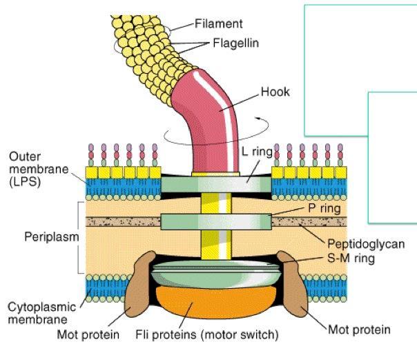
- The basal body attaches the flagellum to the cell wall and plasma membrane.
- It is composed of a small central rod inserted into a series of rings.
- The basal bodies of E. coli and most other typical Gram-negative bacteria have four rings: L, P, MS, and C, which are connected to a central rod (figure 3.42a). The L, P, and MS rings are embedded in the cell envelope, and the C ring is on the cytoplasmic side of the MS ring.
- Typical Grampositive bacteria have only two rings: an inner ring connected to the plasma membrane and an outer one probably attached to the peptidoglycan.

Hook:
- The hook is present outside the cell wall and connects filament to the basal body.
- It consists of different proteins.
- The hook in Gram-positive bacteria is slightly longer than the Gram-negative bacteria.
- it connects filament to the motor protein in the base.
- it is the wider region at the base of filament
Filament or shaft:
- The outermost long region of the flagellum is called filament or shaft
- It has a constant diameter and is made up of globular proteins, the flagellin.the flagellins are arranged in several chains that intertwine and form a helix around a hollow core
- The proteins of flagella act to identify certain pathogenic bacteria unlike eukaryotes, the filaments are not covered by a membrane or sheath.

Type of Flagella
Bacterial species often differ in their patterns of flagella distribution, and these patterns are useful in identifying bacteria.
1. Monotrichous:
- bacteria (trichous means hair) have one flagellum; if it is located at an end, it is said to be a polar flagellum.
- Examples; Vibrio cholera, Pseudomonas aerogenosa
2. Lophotrichous
- bacteria (lopho means tuft) have a cluster of flagella at one or both ends.
- Example: Pseudomanas fluroscence
3. Amphitrichous:
- bacteria (amphi means on bothsides) have a single flagellum at each pole.
- Example; Aquaspirillium
4. Peritrichous:
- Flagella are spread evenly over the whole surface of peritrichous (peri means around) bacteria.
- Example; E.coli, Salmonella, Klebsiella
5. Atrichous:
- They don't contain any flagella.
- Example; Shigella

Functions of Flagella
- The primary function is they help in Movements of unicellular and multi-cellular organisms.
- They also help in Sensatio.
- Signal transduction
- They help in adhesion of organism.
- For cells anchored in a tissue, like the epithelial cells lining our air passages, this moves liquid over the surface of the cell (e.g., driving particle-laden mucus toward the throat).
- Flagella are generally accepted as being important virulence factors


Comments
Post a Comment