This website provide you important notes on Biochemistry,Immunology,Cell Biology,Bacteriology.
Capsule Staining- Principle, Reagents, Procedure and Result
Get link
Facebook
X
Pinterest
Email
Other Apps
The main purpose of capsule stain is to distinguish capsular material from the bacterial cell. A capsule is
a gelatinous outer layer secreted by bacterial cell and that surrounds
and adheres to the cell wall. Most capsules are composed of
polysaccharides, but some are composed of polypeptides. The capsule differs from the slime layer that
most bacterial cells produce in that it is a thick, detectable,
discrete layer outside the cell wall. The capsule stain employs an
acidic stain and a basic stain to detect capsule production.
Principle of Capsule Staining
Capsules stain very poorly with reagents used in simple staining and a
capsule stain can be, depending on the method, a misnomer because the
capsule may or may not be stained.
Negative staining methods contrast a translucent, darker colored,
background with stained cells but an unstained capsule. The
background is formed with india ink or nigrosin or congo red. India ink is difficult to obtain nowadays; however, nigrosin is easily acquired.
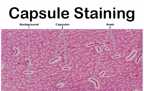
A positive capsule stain requires a mordant that precipitates the capsule. By counterstaining with dyes like crystal violet or methylene blue, bacterial cell wall takes up the dye. Capsules appear colourless with stained cells against dark background.
Capsules are fragile and can be diminished, desiccated, distorted, or
destroyed by heating. A drop of serum can be used during smearing to
enhance the size of the capsule and make it more easily observed with a
typical compound light microscope.
Reagents used for Capsule Staining
Crystal Violet (1%)
Crystal Violet (85% dye content) = 1 gm
Distilled Water = 100 ml Nigrosin
Nigrosine, water soluble = 10 gm
Distilled Water = 100 ml
Procedure of Capsule Staining
Place a small drop of a negative stain (India Ink, Congo Red, Nigrosin, or Eosin) on the slide. Congo Red is easier to see, but it does not work well with some strains. India Ink generally works, but it has tiny particles that display Brownian motion that must be differentiated from your bacteria.Nigrosin may need to be kept very thin or diluted.
Using sterile technique, add a loopful of bacterial culture to slide, smearing it in the dye.
Use the other slide to drag the ink-cell mixture into a thin film along the first slide and let stand for 5-7 minutes.
Allow to air dry (do not heat fix).
Flood the smear with crystal violet stain (this will stain the cells but not the capsules) for about 1 minutes. Drain the crystal violet by tilting the slide at a 45 degree angle and let stain run off until it air dries .
Examine
the smear microscopically (100X) for the presence of encapsulated cells
as indicated by clear zones surrounding the cells.
Result of Capsule Staining
Capsule: Clear halos zone against dark background No Capsule: No Clear halos zone
Examples of Capsule Positive and Negative
Positive Bacillus anthracis, Klebsiella pneumoniae, Streptococcus pneumonia Neisseria meningitidis Clostridiumspp, Rhizaobium spp, etc. NegativeNeisseria gonorrhoreae, etc.
Mneomonics to remember capsulated bacteria- Some Killers Have Pretty Nice Capsule
E. coli is a sort of microorganisms that regularly live in the digestion tracts of individuals and creatures. Be that as it may, a few sorts of E. coli, especially E. coli O157:H7, can cause intestinal disease. E. coli O157:H7 and different strains that cause intestinal disorder are called Shiga poison creating E. coli (STEC) after the poison that they produce. Manifestations of intestinal disease incorporate the runs, stomach torment, and fever. More serious cases can prompt bleeding the runs, lack of hydration, or even kidney disappointment. Individuals with debilitated safe frameworks, pregnant ladies, little youngsters, and more seasoned grown-ups are at expanded danger for building up these difficulties. Most intestinal diseases are brought about by sullied food or water. Legitimate food planning and great cleanliness can incredibly diminish your odds of building up an intestinal disease. Most instances of intestinal E. coli disease can be treated at h...
Complement Pathways: Types, Functions and Regulation Complement was discovered by Jules Bordet as a heat-labile component of normal plasma that causes the opsonisation and killing of bacteria. It helps antibodies and phagocytic cells to clear pathogens and damaged cells; promote inflammation and attack pathogen’s plasma membrane. Proteins that take part in the complement system are called complements that collectively work as a biological cascade The complement system refers to a series of >20 proteins, circulating in the blood and tissue fluids. Most of the proteins are normally inactive, but in response to the recognition of molecular components of microorganisms they become sequentially activated in an enzyme cascade – the activation of one protein enzymatically cleaves and activates the next protein in the cascade. Complements are mainly denoted by the capital letter C with numbers; like, C1, C2, C3, and so on. Some have only alphabet, like, B , D . Some are simpl...
The runs, or watery stools, can be humiliating and strike at the very least occasions, for example, during an excursion or unique occasion. In any case, while loose bowels frequently enhances its own inside a few days, a couple of cures can help advance firmer stools quicker. Continue perusing to find out around five quick acting strategies, alongside what ordinarily causes looseness of the bowels and anticipation tips. 1. Hostile to diarrheal prescription A few people consider looseness of the bowels to be just a mellow disturbance and let it run its course, particularly since certain sessions endures under 24 hours. You may remain nearby to home or a washroom, and burden up on liquids and electrolytes to forestall drying out. Yet, consider the possibility that you can't remain at home. For this situation, taking an enemy of diarrheal medicine may decrease or totally dispense with free stools after the main portion. Search for over-the-counter ...


Comments
Post a Comment